Speaker
Description
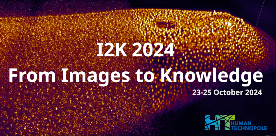
A longstanding quest in biology is to understand how the fertilized egg gives rise to multiple cell types organized in tissues and organs of specialized shape, size and function to suit the lifestyle of multicellular organisms. The integration of molecular, microscopy and image analysis techniques increasingly enables scientists to quantify developmental processes in live developing embryos with single-cell resolution. Here, crustacean Parhyale hawaiensis embryos with fluorescently labeled nuclei were imaged for 3.5 days using a SiMView light-sheet fluorescence microscope. A combination of Matlab and Fiji/ImageJ pipelines were used to produce spatiotemporally registered and fused 4D (3D+time) volumes of each developing embryo. Complete cell lineages, represented as tree graphs, were reconstructed with the Mastodon plugin of Fiji/ImageJ. A new Napari plugin has been developed for pairwise comparisons of cell lineages through the efficient calculation of the (dis)similarity between tree graphs, referred to as the tree edit distance. Visualization of cell lineage comparisons with heat maps and clones on digital embryos paves the way for systems-wide studies of the levels of stereotypy and variability of (sub)lineages within embryos, across embryos and conditions.
| Authors | Giannis Liaskas*, John Rallis, Dimitris Papageorgiou, Evangelia Stamataki, William C. Lemon, Philipp J. Keller, Léo Guignard, Anastasios Pavlopoulos |
|---|---|
| Keywords | light-sheet fluorescence microscopy, nuclear segmentation and tracking, cell lineages, Napari, tree edit distance |

